
The Third Nerve is very important clinically and can be of significant clinical value.
Diagram of the 3rd Nerve Nucleus

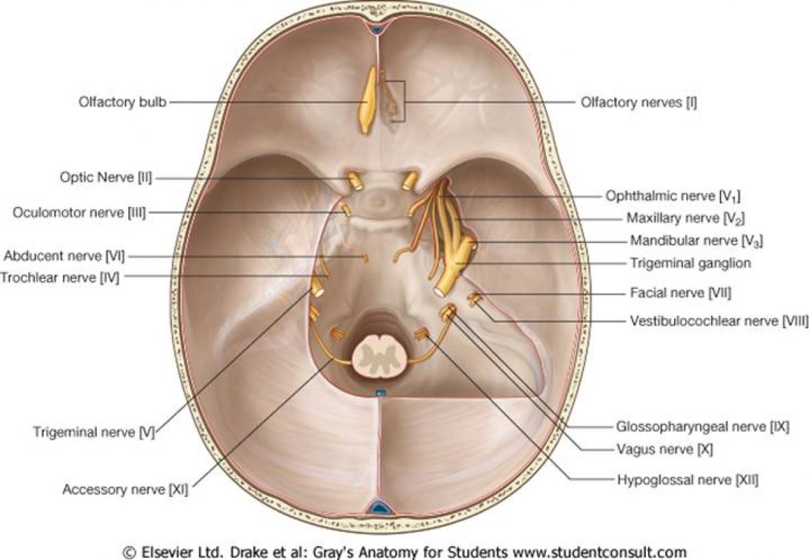
The Route of the Third Nerve is important as this can be useful in trying to diagnose lesions:
- The Oculomotor Nerve Nucleus lies in the Midbrain at the level of the Superior Collicus
- It passes anteriorly into the interpeduncular cistern
- It runs closely alongside the Posterior Communicating Artery
- It runs alongside the tentorium coursing with the Posterior Cerebral Artery
- It enters the Cavernous sinus where it lies in the lateral wall
- It runs in the superior orbital fissure to supply the Levator Palpaebra Superiorus, Sphincter Pupillae (parasympathetic nervous fibres), all extra-ocular muscles bar the lateral rectus and superior oblique muscle
Relationship between the P.Comm Artery and 3rd Nerve

A clinical point the parasympathetic nerve fibres supplying the sphincter pupillae run on the outside of the 3rd nerve and hence are subject to clinical compression. The significant areas of compression include by aneurysms via the P.Comm Aneurysm/Basilar Tip Aneurysm and via uncal herniation via compression of the third nerve at the tentorium.
Those patients with compressive lesions will have pupillary dilation in comparison to non-compressive lesions. This is a key feature of a compressive 3rd nerve palsy.
Causes:
- Uncal Herniation
- Aneurysmal compression
- Diabetes
- Multiple Sclerosis
- Vasculitis
Clinical Features:
- Ptosis (due to Levator Palpabrae Superiorus)
- Pupil ‘down and out’ due to unopposed action of the superior oblique and lateral rectus muscle
- Pupillary dilation
